ФОРМИРОВАНИЕ КИШЕЧНИКА
ТОНКИЙ КИШЕЧНИК
В течение 10-й недели кишечник возвращается в брюшную полость, происходит исчезновение грыжи средней кишки. Тонкий кишечник, образуемый краниальным рукавом петли средней кишки, возвращается первым, проходя позади верхней брыжеечной артерии и занимая центральную часть брюшной полости (Рис 1 А ) , то происходит дальнейший поворот петли средней кишки еще на 90 градусов против часовой стрелки, а в целом на 180 градусов по отношению к исходному положению.
Когда из пупочного канатика возвращается и толстая кишка, то происходит последний поворот еще на 90 градусов, всего же на 270 градусов (Рис.1 Б). Слепая кишка, составляющая самую широкую часть кишечника возвращается последней. Она занимает правую сторону брюшной полости, под правой долей печени, которая в это время достигает нижней части поясничной области.
(Рис 1 В ) , 
Рис.1. Схематическое изображение последовательных этапов поворота средней кишки. А - 10 недель. Б - 11 недель (поворот на 90 градусов). В - 30 недель . Внизу показаны поперечные срезы через петлю средней кишки, через исходный краниальный и каудальный рукава петли средней кишки.
1 - дивертикул слепой кишки, 2 - первоначальное положение желточного канатика, 3 - пупок, 4 - аорта, 5 - верхняя брыжеечная артерия, 6 - малый мешок, 7 - большой сальник, 8 - малый сальник, 9 - отверстие epiploic, 10 - печеночный изгиб, 11 - слепая кишка, 12 - большой сальник, 13 - селезеночный изгиб, 14 - тонкая кишка, 15 - толстая кишка, 16 - аппендикс Fig. 2. Schematic drawings illustrating the rotation of the midgut, as seen from the left. A - 10 week, Б - 11 week, B - 30 week . Transverse section through the midgut loop (down), illustrating the initial relationship of the limbs of the midgut loop to the artery. Б - 90-degree counerclockwise rotation (down) that carries the cranial limb of the midgut to the right.
1 - cecal diverticul, 2 - former site of yolk stalk, 3 - umbilicus, 4 - aorta, 5 - superior mesenteric artery, 6 - lesser sac, 7 - greater omentum, 8 - lesser omentum, 9 - epiploic foramen, 10 - hepatic flexure, 11 - cecum, 12 - greater omentum, 13 - splenic flexure, 14 - smoll intestine, 15 - large intestine, 16 - appendix
Рис.1. Схематическое изображение последовательных этапов поворота средней кишки. А - 10 недель. Б - 11 недель (поворот на 90 градусов). В - 30 недель . Внизу показаны поперечные срезы через петлю средней кишки, через исходный краниальный и каудальный рукава петли средней кишки.
Fig. 2. Schematic drawings illustrating the rotation of the midgut, as seen from the left. A - 10 week, Б - 11 week, B - 30 week . Transverse section through the midgut loop (down), illustrating the initial relationship of the limbs of the midgut loop to the artery. Б - 90-degree counerclockwise rotation (down) that carries the cranial limb of the midgut to the right.
Мышечный слой тонкого кишечника
Ядра гладкомышечных клеток удлиннены и часто дольчатые. Кариоплазма часто содержит 2 ядрышка и богата эухроматином. Цитоплазма мышечных клеток заполнена большим количеством митохондрий и богата цитоплазматическим ретикулемом с грубой поверхностью и содержит аппарат Гольджи. В саркоплазме богатая сеть миофиламент, но электронная плотность их низкая. Плотные области отсутствуют на клеточных мембранах, тогда как имеются плотные тельца, эндоцитотические пузырьки и базальные оболочки. Тела клеток тесно соприкасаются друг с другом, межклеточные пространства очень узкие.
Формируется продольный мышечный слой. Гладкомышечные клетки начинают терять свои эмбриональные характеристики и начинают дифференцироваться во взрослые миоциты (Benedeczky et al., 1993).
Нервные клетки тонкого кишечника
Большая часть нервных клеток все еще представлена нейробластами, которые формируют компактные внутристеночные ганглии (парасимпатическая нервная система). Ядра их богаты как гетеро-, так и эухроматином. Перикарион обычно узкий и содержит большое количество свободных рибосом, мало митохондрий и трубочек грубого эндоплазматического ретикулема. Цитоплазма Шванновских клеток электрон прозрачна. Среди нейрональных клеток обнаруживаются простые нейропили. Нервнвые отростки содержат хорошо развитую систему микротрубочек, сферические пузырьки. В некоторыых нервных отростках обнаруживается небольшое количество митохондрий и свободныхъ рибосом. Формируются синаптические контакты между нейрональными элементами. Пузырьков на окончаниях аксонов довольно мало. Нейромышечные контакты встречаются часто. Некоторые нервные отростки формируют сплетения и аксолемма находится в тесном контакте с сарколеммой (Benedeczky et al., 1993).
Как и в парасимпатической симпатической нервной системе предшествует развитие преганглиолярных нейронов, в данном случае клеток боковых рогов спинного мозга. У зародышей 11-12 недель развития эти клетки обнаруживают первые признаки дифференцировки - рост ядра, появление отростков, усложнение нейрофибриллярного аппарата и возрастание его сродства к серебру, утрата равномерного распределения ДНК хромосом по ядру, перинуклеарное развитие нисслевких телец, накопление в ядрышке основных белков и т.д.
ТОЛСТЫЙ КИШЕЧНИК
На 4-м месяце в слизхистой оболочке толстой кишки начинают формироваться крипты и выросты слизхистой оболочки, похожие на ворсинки тонкой кишки. Последние существуют почти до конца внутриутробной жизни, затем сглаживаются и исчезают. Одновременно с образованием крипт в их эпителиальной выстилке появляется множество бокаловидных клеток, вырабатывающих слизь.
Прямая кишка
Все процессы дифференцировки слизистой оболочки толстой кишки, связанные с образованием крипт и ворсинок, появлением бокаловидных клеток, раньше всего начинают и достигают наибольшего развития в области прямой кишки, а затем уже становятся заметными в других отделах толстого кишечника
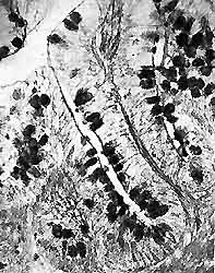
Клетки бокаловидного эпителия в криптах прямой кишки. Плод длиной 68 мм.
ТОЛСТЫЙ КИШЕЧНИК
На 4-м месяце в слизхистой оболочке толстой кишки начинают формироваться крипты и выросты слизхистой оболочки, похожие на ворсинки тонкой кишки. Последние существуют почти до конца внутриутробной жизни, затем сглаживаются и исчезают. Одновременно с образованием крипт в их эпителиальной выстилке появляется множество бокаловидных клеток, вырабатывающих слизь.
Прямая кишка
Все процессы дифференцировки слизистой оболочки толстой кишки, связанные с образованием крипт и ворсинок, появлением бокаловидных клеток, раньше всего начинают и достигают наибольшего развития в области прямой кишки, а затем уже становятся заметными в других отделах толстого кишечника
Клетки бокаловидного эпителия в криптах прямой кишки. Плод длиной 68 мм.
Известно, что появление выраженной реакции сокращающегося кишечника человеческого зародыша на пилокарпин и адреналин совпадают по времени (10-12 неделя) с началом процессов дифференцировки нервноклеточных элементов ауэрбаховского сплетения и образования связей их отростков с окружающими тканями. Для нормального развития нервной клетки необходимы раздражения с периферии.