Expression of FoxP2 during zebrafish development and in the adult brain | |
|
|
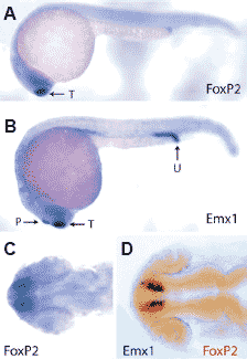
 Fig. 2. (Left) Expression of FoxP2 during
zebrafish embryogenesis. (A) Lateral view of
in situ hybridization of FoxP2 probe to a 20-
somite zebrafish embryo. The expression is in
the dorsal telencephalon (arrow). (B) Lateral
view of in situ hybridization of Emx1 probe to a
20-somite zebrafish embryo. Expression is in the dorsal telencephalon (T), pineal gland (P) and the urogenital opening (U). (C) Dorsal view of the head region from a 20-somite zebrafish embryo
hybridized with a FoxP2 probe. (D) Double in situ hybridization of a FoxP2 probe (red) and Emx1 (black) to the head region of a 20-somite zebrafish
embryo. Dorsal view. Fig. 2. (Left) Expression of FoxP2 during
zebrafish embryogenesis. (A) Lateral view of
in situ hybridization of FoxP2 probe to a 20-
somite zebrafish embryo. The expression is in
the dorsal telencephalon (arrow). (B) Lateral
view of in situ hybridization of Emx1 probe to a
20-somite zebrafish embryo. Expression is in the dorsal telencephalon (T), pineal gland (P) and the urogenital opening (U). (C) Dorsal view of the head region from a 20-somite zebrafish embryo
hybridized with a FoxP2 probe. (D) Double in situ hybridization of a FoxP2 probe (red) and Emx1 (black) to the head region of a 20-somite zebrafish
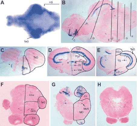
embryo. Dorsal view. Fox (forkhead) гены кодируют транскрипционные факторы, которые играют важные роли в регуляции формирования эмбрионального паттерна, а также экспрессии ткане-специфических генов. Мутации в гене FOXP2 человека вызывают аномалии речи. Описывается структура и паттерн экспрессии FoxP2 рыбок данио. У рыбок данио этот ген впервые обнаруживает экспрессию на ст. 20 сомитов в презумптивном telencephalon.На этой стадии обнаруживается существенное перекрывание экспрессии FoxP2 с экспрессией emx гомеобоксных генов. Однако, в противоположность emx1, FoxP2 не экспрессируется в шишковидной железе или в пронефрических протоках. После 72 ч развития экспрессия FoxP2 у рыбок данио становится более сложной в головном мозге. Развивающийся зрительный tectum становится главной областью экспрессии FoxP2. В головном мозге взрослых особей наивысшие концентрации транскриптов FoxP2 обнаруживаются в optic tectum. В мозжечке только каудальные доли обнаруживают высокие уровни экспрессии Foxp2. Эти области соответствуют vestibulocerebellum у млекопитающих. Некоторые др. области головного мозга также обнаруживают высокие уровни экспрессии Foxp2.  Fig. 3. (Right) Expression of FoxP2 in the zebrafish brain. (A) Dorsal view of in situ hybridization of FoxP2 probe to the isolated brain from a 7 dayold zebrafish. The isolated brain was opened along its dorsal axis and flattened. Anterior is to the left. (B) Sagittal section of a brain from a 3 months old zebrafish hybridized with a FoxP2 probe. Vertical lines indicate the positions of cross sections in images (C - H). Cross sections, hybridized with a FoxP2 probe, through the (C) telencephalon, (D)optic tectum, (E) optic tectum, cerebellum and hypothalamus, (F) cerebellum, (G) caudal lobe of the cerebellum and the medulla oblongata. Arrow in (F) indicates the expression in the superior reticular formation. Arrow in (G) indicates expression in the medial octavolateralis nucleus. (H) A section caudal to (G) shows no expression of FoxP2. Abbreviations: Cc, cerebellar crest; CaC, caudal lobe of cerebellum; CCe, corpus cerebelli; DIL, diffuse nucleus of the inferior hypothalamic lobe; Dt, dorsal thalamus; HB, hindbrain; HT, hypothalamus; MO, medulla oblongata; MoN, medial octavolateralis nucleus; PgZ, periventicular gray zone of the optic tectum; Po, preoptic area; Pvpt, periventricular pretectum; SRN, superior reticular nucleus; TeO, optic tectum; TS, torus semicircularis; Vpt, ventral posterior tuberculum. |