ФОРМИРОВАНИЕ ГРАНИЦ
Kenneth D Irvine
Current Opinion in Genetics & Development 1999, 9, No. 4:434-441.
- A–P—anterior–posterior;
D–V—dorsal–ventral;
Dpp—Decapentaplegic;
Hh—Hedgehog;
L-Fng—Lunatic Fringe;
R-Fng—Radical Fringe.
В исследованиях на крыльях Drosophila было установлено, что Fringe ингибирует эффективность Notch лиганда Serrate и потенцирует эффективность Notch лиганда Delta [8] [9]. Fringe обычно экспрессируется дорсальными клетками [11], в результате происходит активация Notch вдоль D–V границы крыла. Гомолог Fringe у позвоночных, Radical Fringe (R–Fng), экспрессируется специфически дорсальными клетками в развивающемся зачатке конечности кур и его функция аналогична его роли у Drosophila [12] [13].
Fleming RJ, Gu Y, Hukriede NA:
Serrate-mediated activation of Notch is specifically blocked by the product of the gene fringe in the dorsal compartment of the Drosophila wing imaginal disc.
Development 1997, 124: 2973–2981.
Panin VM, Papayannopoulos V, Wilson R, Irvine KD:
Fringe modulates Notch–ligand interactions.
Nature 1997, 387: 908–912.
de Celis JF, Bray S:
Feed-back mechanisms affecting Notch activation at the dorsoventral boundary in the Drosophila wing.
Development 1997, 124: 3241–3251.
Irvine KD, Wieschaus E:
fringe, a boundary-specific signaling molecule, mediates interactions between dorsal and ventral cells during Drosophila wing development.
Cell 1994, 79: 595–606.
Rodriguez-Esteban C, Schwabe JWR, De La Pena J, Foys B, Eshelman B, Izpisua Belmonte JC:
Radical fringe positions the apical ectodermal ridge at the dorsal-ventral boundary of the vertebrate limb.
Nature 1997, 386: 360–366.
Laufer E, Dahn R, Orozco OE, Yeo C-Y, Pisenti J, Henrique D, Abbott UK, Fallon JF, Tabin C:
Expression of Radical fringe in limb-bud ectoderm regulates apical ectodermal ridge formation.
Nature 1997, 386: 366–373.
Hukriede NA, Gu Y, Flemming RJ:
A dominant-negative form of Serrate acts as a general antagonist of Notch activation.
Development 1997, 124: 3427–3437.
Sun X, Artavanis-Tsakonas S:
Secreted forms of DELTA and SERRATE define antagonists of Notch activation.
Development 1997, 124: 3439–3448.
Klueg KM, Parody TR, Muskavitch MA:
Complex proteolytic processing acts on Delta, a transmembrane ligand for Notch, during Drosophila development.
Mol Biol Cell 1998, 9: 1709–1723.
• Qi H, Rand MD, Wu X, Sestan N, Wang W, Rakic P, Xu T, Artavanis-Tsakonas S:
Processing of the Notch ligand Delta by the metalloprotease Kuzbanian.
Science 1999, 283: 91–94.
• Neumann CJ, Cohen SM:
Boundary formation in Drosophila wing: Notch activity attenuated by the POU protein Nubbin.
Science 1998, 281: 409–413.
- Ng M, Diaz-Benjumea FJ, Cohen SM:
nubbin encodes a POU-domain protein required for proximal–distal patterning in the Drosophila wing.
Development 1995, 121: 589–599.
- • Moran JL, Levorse JM, Vogt TF:
Limbs and beyond the Radical Fringe.
Nature 1999, 399: 742–743.
- • Zhang N, Gridley T:
Defects in somite formation in lunatic fringe-deficient mice.
Nature 1998, 394: 374–377.
- • Evrard YA, Lun Y, Aulehla A, Gan L, Johnson RL:
lunatic fringe is an essential mediator of somite segmentation and patterning.
Nature 1998, 394: 377–381.
- Christen B, Slack JMW:
All limbs are not the same.
Nature 1998, 395: 230–231.
- • Papayannopoulos V, Tomlinson A, Panin VM, Rauskolb C, Irvine KD:
Dorsal-ventral signaling in the Drosophila eye.
Science 1998, 281: 2031–2034.
- Bachmann A, Knust E:
Dissection of cis-regulatory elements of the Drosophila gene Serrate.
Dev Genes Evol 1998, 208: 346–351.
- • Dominguez M, de Celis JF:
A dorsal/ventral boundary established by Notch controls growth and polarity in the Drosophila eye.
Nature 1998, 396: 276–278.
- • Cho K-O, Choi K-W:
Fringe is essential for mirror symmetry and morphogenesis in the Drosophila eye.
Nature 1998, 396: 272–276.
- Brodsky MH, Steller H:
Positional information along the dorsal-ventral axis of the Drosophila eye: graded expression of the four-jointed gene.
Dev Biol 1996, 173: 428–446.
- Villano JL, Katz FN:
four-jointed is required for intermediate growth in the proximal-distal axis in Drosophila.
Development 1995, 121: 2767–2777.
- Sun YH, Tsai C-J, Green MM, Chao J-L, Yu C-T, Jaw TJ, Yeh J-Y, Bolshakov VN:
white as a reporter gene to detect transcriptional silencers specifying position-specific gene expression during Drosophila melanogaster eye development.
Genetics 1995, 141: 1075–1086.
- Heberlein U, Borod ER, Chanut FA:
Dorsoventral patterning in the Drosophila retina by wingless.
Development 1998, 125: 567–577.
- Williams JA, Paddock SW, Carroll SB:
Pattern formation in a secondary field: a hierarchy of regulatory genes subdivides the developing Drosophila wing disc into discrete subregions.
Development 1993, 117: 571–584.
- Gomez-Skarmeta JL, del Corral RD, Calle-Mustienes dl, Ferre MD, Modolell J:
Araucan and caupolican, two members of the novel Iroquois complex, encode homeo-proteins that control proneural and vein-forming genes.
Cell 1996, 85: 95–105.
- McNeill H, Yang C-H, Brodsky M, Ungos J, Simon MA:
mirror encodes a novel PBX-class homeoprotein that functions in the definition of the dorsal-ventral border in the Drosophila eye.
Genes Dev 1997, 11: 1073–1082.
- Netter S, Fauvarque MO, Diez del Corral R, Dura JM, Coen D:
white+ transgene insertions presenting a dorsal/ventral pattern define a single cluster of homeobox genes that is silenced by the polycomb-group proteins in Drosophila melanogaster.
Genetics 1998, 149: 257–275.
- • Rauskolb C, Irvine KD:
Notch-mediated segmentation and growth control of the Drosophila leg.
Dev Biol 1999, 210: 339–350.
- • de Celis JF, Tyler DM, de Celis J, Bray SJ:
Notch signaling mediates segmentation of the Drosophila leg.
Development 1998, 125: 4617–4626.
- Speicher SA, Thomas U, Hinz U, Knust E:
The Serrate locus of Drosophila and its role in morphogenesis of the wing imaginal discs: control of cell proliferation.
Development 1994, 120: 535–544.
- Shellenbarger DL, Mohler JD:
Temperature-sensitive periods and autonomy of pleiotropic effects of I(1)Nts1, a conditional Notch lethal in Drosophila.
Dev Biol 1978, 62: 432–446.
- Parody TR, Muskavitch MAT:
The pleiotropic function of Delta during postembryonic development of Drosophila melanogaster.
Genetics 1993, 135: 527–539.
- Basler K, Struhl G:
Compartment boundaries and the control of Drosophila limb pattern by hedgehog protein.
Nature 1994, 368: 208–214.
- Diaz-Benjumea FJ, Cohen B, Cohen SM:
Cell interaction between compartments establishes the proximal-distal axis of Drosophila legs.
Nature 1994, 372: 175–179.
- Struhl G, Basler K:
Organizing activity of wingless protein in Drosophila.
Cell 1993, 72: 527–540.
- Lecuit T, Cohen SM:
Proximal-distal axis formation in the Drosophila leg.
Nature 1997, 388: 139–145.
- Abu-Shaar M, Mann RS:
Generation of multiple antagonistic domains along the proximodistal axis during Drosophila leg development.
Development 1998, 125: 3821–3830.
- Wu J, Cohen SM:
Proximodistal axis formation in the Drosophila leg: subdivision into proximal and distal domains by Homothorax and Distal-less.
Development 1999, 126: 109–117.
- Pankratz M, Jackle H: Blastoderm segmentation. In The Development of Drosophila melanogaster. Edited by Bate M, Martinez Arias A. Cold Spring Harbor, New York: Cold Spring Harbor Laboratory Press, 1993, 467–516.
- Conlon RA, Reaume AG, Rossant J:
Notch1 is required for the coordinate segmentation of somites.
Development 1995, 121: 1533–1545.
- Jiang YJ, Smithers L, Lewis J:
Vertebrate segmentation: the clock is linked to Notch signalling.
Curr Biol 1998, 8: R868–R871.
- McGrew MJ, Pourquie O:
Somitogenesis: segmenting a vertebrate.
Curr Opin Genet Dev 1998, 8: 487–493.
- Keynes RJ, Stern CD:
Mechanisms of vertebrate segmentation.
Development 1988, 103: 413–429.
- Johnston SH, Rauskolb C, Wilson R, Prabhakaran B, Irvine KD, Vogt TF:
A family of mammalian Fringe genes implicated in boundary determination and the Notch pathway.
Development 1997, 124: 2245–2254.
- Cohen B, Bashirullah A, Dagnino L, Campbell C, Fisher WW, Leow CC, Whiting E, Ryan D, Zinyk D, Boulianne G et al.:
Fringe boundaries coincide with Notch-dependent patterning centres in mammals and alter Notch-dependent development in Drosophila.
Nat Genet 1997, 16: 283–288.
- de la Pompa JL, Wakeham A, Correia KM, Samper E, Brown S, Aguilera RJ, Nakano T, Honjo T, Mak TW, Rossant J, Conlon RA:
Conservation of the Notch signalling pathway in mammalian neurogenesis.
Development 1997, 124: 1139–1148.
- Kusumi K, Sun ES, Kerrebrock AW, Bronson RT, Chi DC, Bulotsky MS, Spencer JB, Birren BW, Frankel WN, Lander ES:
The mouse pudgy mutation disrupts Delta homologue DII3 and initiation of early somite boundaries.
Nat Genet 1998, 19: 274–278.
- • Forsberg H, Crozet F, Brown NA:
Waves of mouse Lunatic fringe expression, in four-hour cycles at two-hour intervals, precede somite boundary formation.
Curr Biol 1998, 8: 1027–1030.
- • McGrew MJ, Dale JK, Fraboulet S, Pourquie O:
The lunatic fringe gene is a target of the molecular clock linked to somite segmentation in avian embryos.
Curr Biol 1998, 8: 979–982.
- • Aulehla A, Johnson RL:
Dynamic expression of lunatic fringe suggests a link between notch signaling and an autonomous cellular oscillator driving somite segmentation.
Dev Biol 1999, 207: 49–61.
- Palmeirim I, Henriques D, Ish-Horowicz D, Pourquie O:
Avian hairy gene expression identifies a molecular clock linked to vertebrate segmentation and somitogenesis.
Cell 1997, 91: 639–648.
- Micchelli CA, Rulifson EJ, Blair SS:
The function and regulation of cut expression on the wing margin of Drosophila: Notch, Wingless and a dominant negative role for Delta and Serrate.
Development 1997, 124: 1485–1495.
- Holland LZ, Kene M, Williams NA, Holland ND:
Sequence and embryonic expression of the amphioxus engrailed gene (AmphiEn): the metameric pattern of transcription resembles that of its segment polarity homolog in Drosophila.
Development 1997, 124: 1723–1732.
- Wehrli M, Tomlinson A:
Independent regulation of anterior/posterior and equatorial/polar polarity in the Drosophila eye; evidence for the involvement of Wnt signaling in the equatorial/polar axis.
Development 1998, 125: 1421–1432.
- Bettenhausen B, Hrabe de Angelis M, Simon D, Guenet J, Gossler A:
Transient and restricted expression during mouse embryogenesis of DII1, a murine gene closely related to Drosophila Delta.
Development 1995, 121: 2407–2418.
- Dunwoodie SL, Henrique D, Harrison SM, Beddington RS:
Mouse DII3: a novel divergent Delta gene which may complement the function of other Delta homologues during early pattern formation in the mouse embryo.
Development 1997, 124: 3065–3076.
- Zeilder MP, Perrimon N, Strutt DI:
Polarity determination in the Drosophila eye: a novel role for Unpaired and JAK/STAT signaling.
Genes Dev 1999, 13: 1342–1353.
- Bishop SA, Klein T, Martinez Arias A, Couso JP:
Composite signaling from Serrate and Delta establish leg segments in Drosophila through Notch.
Development 1999, 126: 2993–3003.
- Jen W-C, Gawantka V, Pollet N, Niehrs C, Kintner C:
Periodic repression of Notch pathway genes governs the segmentation of Xenopus embryos.
Genes Dev 1999, 13: 1486–1499.
- Takke C, Campos-Ortega JA:
her1, a zebrafish pair-rule like gene, acts downstream of notch signalling to control somite development.
Development 1999, 126: 3005–3014.
- del Barco Barrantes I, Elia AJ, Wunsch K, Hrabe De Angelis M, Mak TW, Rossant J, Conlon RA, Gossler A, de la Pompa JL:
Interaction between Notch signaling and Lunatic fringe during somite boundary formation in the mouse.
Curr Biol 1999, 9: 470–480.
- Ng M, Diaz-Benjumea FJ, Cohen SM:
Dorsal–ventral limb borders
Имеются указания на то, что существуют механизмы эндогенного процессинга лигандов [15] [16] [17•]которые могут генерировать растворимые формы Delta, способные активировать Notch [17•]. Более того, исследования функции гена nubbin указывают на то, что потенциал ширины полосы активации Notch много шире, чем предполагалось ранее[18•]. nubbin кодирует POU доменовый белок, который экспрессируется во всем зачатке крыла [19]. Экспрессия по крайней мере двух Notch target генов вдольD–V границы негативно регулируется nubbin [18•]. Если активность nubbin удалена, то экспрессия этих Notch генов-мишеней может обнаруживаться на расстоянии в 10 клеток от D–V границы. У кур выявляется сходная роль R-Fng [12] [13]; [20•], но у млекопитающих и лягушек роль Fringe генов в развитии конечностей установить пока не удается [21•] [22•] (T Vogt, personal communication). [23].
Specification of dorsal–ventral midline cells in the Drosophila eye
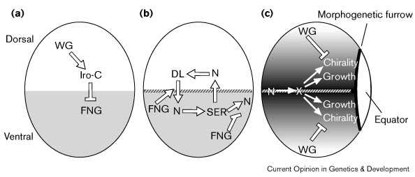
Хотя клетки D–среденей линии не отличаются по морфологии, идентифицированы некоторые гены, экспрессирующиеся преимущественно вдоль D–V средней линии [24•] [25] [26•] [27•] [28] [29] [30] [31]. Эти молекулярные маркеры D–V средней линии регулируются Notch [24•] [26•] [27•]. Используя эти молекулярные маркеры оказалось возможным показать, что активация Notch позиционируется с помощью Fringe [24•] [27•], который обычно экспрессируется спецефически вентральными клетками глаз [24•] [26•] [27•] (Fig. 1b). Notch лиганды Serrate и Delta экспрессируются преимуществено вдоль D–V средней линии [24•] [25] [26•] [27•] и их экспрессия регулируется активностью Notch [24•] [27•], как и в крыльях (Fig. 1b). Более того, Delta и Serrate обладают дорсальной и вентральной специфичностью своей активности, которая связана с Fringe [24•]. Однако экспрессия Fringe и Notch лигандов инвертирована в отношении D–V оси в глазах и крыльях.
 |
| Рис. 1
Схематическое изображение
последовательных ступеней
формирования D–V паттерна
в глазных имагинальных
дистакх Drosophila;
дорсальная часть вверху, а
передняя - слева. (a)
Детерминация дорсальной и
вентральной судьбы клеток.
Wingless (WG) позитивно
регулирует зеркальную
экспрессию в дорсальных
клетках [31], и,
по-видимому, гены другого
Iroqouis-complex (Iro-C) тоже. Т.к. WG
экспрессируется также в
вентральных клетках, даже
в раннем третьем
личиночном возрасте (не
показано), то должны
существовать
дополнительные факторы
для ограничения
экспрессии Iro-C . Iro-C гены
негативно регулируют Fringe
(FNG, серая заливка) [26•] [27•]. (b) Активация Notch
(N) на срединной линии D–V
(штриховая линия).
Предполагается, что Serrate
(SER) сигналы передаются от
от вентральных клеток
дорсальным, а Delta (DL) от
дорсальных вентральным,
причем FNG играет ключевую
роль в модуляции их
сигнальной активности [9] [24•]. Эти регуляторные ступени не нуждаются в управлении. (c) Активация Notch оказывает дально-действующее влияние как на рост глаз, так и chiralty омматидий[24•] [26•] [27•]. Предполагается, что это происходит с помощью индукции диффузными сигнальными молекулами, Х, которые распространяются от места их синтеза и распределяются градиентно. Сhirality омматидий также зависит от WG; эффекты WG оппозитны тем, что индуцируются Notch [62] Figure 1 Schematic illustrating sequential steps of D–V patterning in the Drosophila eye imaginal disc; dorsal is up and anterior to the left in each panel. (a) Establishment of dorsal and ventral cell fates. Wingless (WG) positively regulates mirror expression in dorsal cells [31], and presumably that of other Iroqouis-complex (Iro-C) genes as well. As WG is also expressed in ventral cells, even at early third instar (not shown), additional factors must also contribute to restricted Iro-C expression. Iro-C genes negatively regulate Fringe (FNG, gray shading) [26•] [27•]. (b) Activation of Notch (N) at the D–V midline (hatched line). Taking into consideration their expression patterns and ability to influence each others' expression when ectopically expressed, Serrate (SER) is hypothesized to signal from ventral cells to dorsal cells, and Delta (DL) from dorsal cells to ventral cells, with FNG playing a key role by modulating their signaling activity [9] [24•]. These regulatory steps need not be direct. (c) Activation of Notch has long-range influences on both eye growth and ommatidial chiralty [24•] [26•] [27•]. This is proposed to occur by induction of a diffusible signaling molecule, X, which spreads from its site of synthesis and becomes distributed in a gradient. Ommatidial chirality is also influenced by WG; the effects of WG are opposite to those induced by Notch activation [62]. |
Самое раннее проявление D–V полярности в крыльях и глазах генерируется с помощью Wingless; однако в крыльях Wingless закладка D–V полярности осуществляется с помощью негативно реглуируемой экспрессии Apterous [32], тогда как в глазах wingless D–V полярность позитивно регулируется экспрессией mirror [31], и возможно другими чоенами комплекса Iroquois (Fig. 1a). Комплекс Iroquois является кластером трех родственных гомеобоксных генов — mirror, araucan and caupolican — которые экспрессируются в виде сходных паттернов во многих ткнях, включая глаза [26•] [33] [34] [35]. Экспрессия комплекса Iroquois комплементарнаfringe в глазах [24•] [26•], и эти гены могут негативно регулировать fringe ([26•] [27•]; H McNeill, personal communication) ограничивая fringe вентральными клетками (Fig. 1a).
Активация Notch вдоль D–V
средней линии глаз играет
критическую роль в
паттернировании и росте глаз [24•] [26•] [27•]. Local
activation of Notch exerts a long-range influence on
ommatidial chirality and is also necessary and sufficient
to promote eye growth (Fig. 1c).
Segmentation
of Drosophila appendages [36•] [37•]. Notch сигналы
создают мноржественные
повторяющиеся границы вдоль
proximal–distal оси конечности .
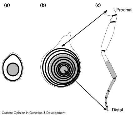
Эктопическая активация Notch может вести к индукции структур эктопических суставов [36•] [37•]. Сегментно повторяющаяся потребность в Notch параллельна сегментно повторяющейся экспрессии его лигандов [25] [36•] [37•], и генов, регулируюемых Notch [36•] [37•]. Notch, Serrate, и Delta функционируют одинаково во время сегментации ног и формирования D–V границы, но имеются и отличия в потребности fringe.
 |
| Figure 2 Progressive segmentation of the Drosophila leg. (a,b) Schematic of leg imaginal disc at (a) early and (b) late third instar. (c) Schematic of adult leg. During metamorphosis, the leg imaginal disc telescopes out such that the center of the leg disc gives rise to the distal tip of the adult leg and more peripheral regions of the disc give rise to regions that are progressively more proximal to the body. Black rings represent regions of Notch activation, which drive leg segmentation and growth [36•] [37•]; gray shading represents expression of Distal-less. (a) Notch activation is detectable in two rings, one proximal and a second that appears at the edge of the Distal-less domain [37•]. This distal ring may correspond to the most distal ring in mature leg discs [37•]. (b) By the end of third instar, rings of Notch activation exist for most or all segment borders. Although all rings are illustrated here, it is not actually possible to visualize them all in one focal plane as a result of the highly folded nature of the disc epithelium. |
Сегментно повторяющаяся
экспрессия Serrate — и возможно fringe
и Delta — регулируется с помощью
Wingless и Decapentaplegic (Dpp) [36•]. Wingless и Dpp
действуют комбинационно
создавая proximal–distal ось
конечности и обеспечивают ее
рост [41] [42] [43]. Они
также регулируют экспрессию
генов таких как Distal-less, dachshund,
и homothorax [44] [45] [46]. Предполагается,
что эти гены экспрессирующиеся
широкими полосами могут
дейстовать комбинационно
создавая отдельные кольца
экспрессиии fringe, Serrate, и Delta [36•] [37•]. (Fig. 2).
Regulation
and function of Lunatic Fringe in vertebrate
somitogenesis Роль сигналов Notch в
сомитогенезе впервые выявлена
с помощью генного таргетинга у
мышей Notch1 [48] и затем
продемонстрирована у кур,
лягушек и рыб (reviewed in [49] [50]). Сомитогнез(reviewed in
[51]) (Fig. 3). Несколько генов
идентифицировано в сомитах.
Эти молекулярные маркеры
сегментации показывают, что
вновь формирующиеся сомиты уже
подразделены на отдельные
передение и задние типы клеток.
 |
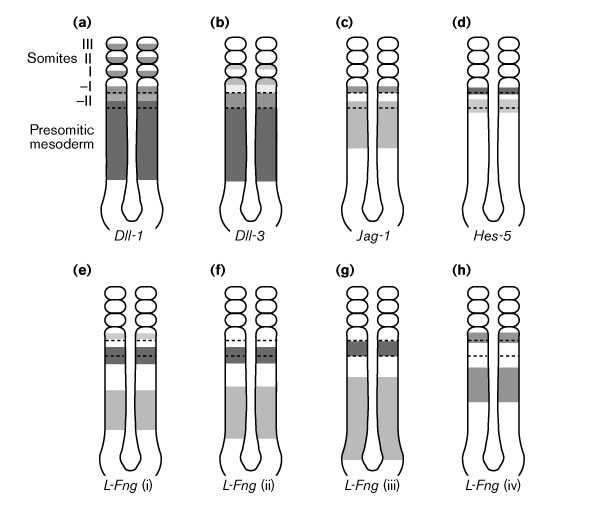
| Figure 3 Expression of L-Fng, Notch ligands, and Hes-5 during mouse somitogenesis. The expression of Notch1 and Notch2 (not shown) is also modulated during somitogenesis. Shading approximates relative levels of expression. Borders between presumptive somites are not identifiable in the presomitic mesoderm, hence the relationships between gene expression patterns and future somite boundaries are approximate. (a) Delta-like 1 (Dll-1) is expressed in the posterior of forming (–I) and newly formed (I) somites [63] [64]. (b) Delta-like 3 (Dll-3) is expressed in the anterior of forming and newly formed somites [64]. (c) Jagged/Serrate-1 (Jag-1) is expressed in the posterior of the forming somite [21•] [53]. (d) Hes-5 is expressed in the posterior or at the posterior border of the forming somite [22•] [54]. (e–h) L-Fng expression is dynamic. (This depiction is adapted largely from [56•].) i–iv represent different steps in a cycle of L-Fng expression. There are some differences between the reported L-Fng expression profiles in mice and chicks and there are also some differences between the profiles described by different groups [52] [53] [56•] [57•] [58•] but, in general, L-Fng expression appears to move in waves towards the anterior (rostral) end of the presomitic mesoderm, and these waves narrow as they move anteriorly. In mice (but not in chicks) the most anterior wave of L-Fng expression fades away before the newly formed somite is morphologically identifiable. |
Нарушение передачи сигналов Notch не приостанавливает сомитогенез, но вызывает дизорганизацию этого процесса, сомитогенез задерживатеся, формируется меньше границ между сомитами и эти границы расположены нерегулярно (reviewed in [49] [50]). Молекулярно, некоторая сегментная периодичность в паттернах экспресссии генов сохраняется, но экспрессия границ более диффузна. Наиболее существенным инициальным дефектом является разрушение A–P компартментализации в формирующихся сомитах: гены, обычно экспрессирующиеся или в передених или в задних клетках, теперь экспрессируются изменчиво по всей ширине сомита. Неясно, существует ли независмая потребность в сигналах Notch как для формирования границ, так и A–P компартментализации, или один из этих фенотипов является вторичным.
Экспрессия в виде полос Lunatic
Fringe (L-Fng) в пресомитной
мезодерме указывает на то. что
он играет важную рольв в
сомитогенезе [52] [53] (Fig. 3) .
Мутанты L-Fng имеют дефекты в A–P
подразделении и в формировании
границ сомитов [21•] [22•]. L-Fng мутанты
также влияют на экспрессию Hes-5 [22•],который
является геном-мишенью для Notch [54].
Нарушение сигнализациии[55] Notch
ведет к более диффузной
экспрессии Notch и Notch лигандов.
Хотя эти нарушения в
экспрессии генов пути Notch могут
быть результатом нарушения
петли обратной связи, сходной с
таковой в крыльях и глазах Drosophila,
однако возможно также, что они
являются вторичным следствием
дефектов формирования сомитов.
L-Fng экспрессия в виде полос
в пресомитной мезодерме на
ранних стадиях сомитогенеза
скорее, чем любого другого
члена пути Notch [52] [53] [56•] [57•] [58•] (Fig. 3).
Более того, L-Fng
регулируется молекулярными
часами, которые функционируют
во время сомитогенеза.
Изучение гена hairy у кур
показало существование этого
аутосомного клеточного
осцилятора, который управляет
циклическими волнами
экспрессии hairy в
пре-сомитной мезодерме [59]. L-Fng
экспрессия испытывает сходные
осциляции [56•]
[57•] [58•], и
так как L-Fng, а не hairy
осциляции зависят от синтеза
белка, кажется наиболее
вероятным, что L-Fng
регулируется с помощью hairy [57•].
Выявляется критическая связь
между клеточным осцилятором и
регуляцией сигналов Notch.
Однако, L-Fng экспрессия в
пресомитной мезодерме
динамична и неясно, какой
аспект паттерна экспрессии
наиболее важен для
сомитогенеза. В формировании
(–I) пре-сомита, L-Fng
экспрессия примерно в половине
сомита, но не в –II пресомите, L-Fng
экспрессия на всю ширину
сомита (Fig. 3). Хотя
экспрессия в половине сомита
может быть сравнена с
постулируемой ролью A–P
компартментализации, L-Fng
экспрессируется в переденей
половине –I сомита кур [22•] [57•], и в
задней половине –I сомита у
мышей [52]
[56•], и,
по-видмому, мало вероятно, что
мыши и куры используют
различные механизмы A–P
компартментализации.
Conclusions
fringe модулирует передачу сигналов Notch так, что активация Notch оказывается позиционированной вдоль границы экспрессии fringe.