Посещений:
ПРЕКАРДИАЛЬНЫЕ КЛЕТКИ ПЕРВИЧНОЙ ПОЛОСКИ

Детерминация, Перемещения

Movement and commitment of primitive streak precardiac cells during cardiogenesis
CARMEN LOPEZ-SANCHEZ, NATIVIDAD GARCIA-MASA, CARLOS M. GANAN and VIRGINIO GARCIA-MARTINEZ
Int. J. Dev. Biol. 53: 1445-1455 (2009) doi: 10.1387/ijdb.072417cl

ABSTRACT Fate maps are required to address questions about the commitment and differentiation of precardiac cells. Here, we report a detailed study of the precardiac cells located at the level of the primitive streak, employing different experiments with a variety of techniques combining double transplantations, microinjections and immunocytochemistry. Most cells of the more rostral segments of the primitive streak were found to contribute cells to the endodermal layer, adjacent to precardiac mesodermal cells of the heart forming region whose provenance was in the immediately more caudal segments of the primitive streak. We established a close spatiotemporal relationship between the two cell layers and the expression of their specific cardiac markers ( cNkx-2.5, Bmp2, Cripto, Usmaar, dHand, GATA4, Pitx2, Hex, Fgf8, AMHC1 and VMHC1). We also analyzed the ability of precardiac cells to differentiate when they are transplanted to ectopic locations or are subjected to the influence of the organizer. We propose that the precardiac cells of the primitive streak form at least two groups with different significance. One, regulated by mediation of the organizer, is located preferentially in the more rostral region of the primitive streak. It consists of the prospective cells of the endoderm layer, with a hierarchic pattern of expression of different genes characterized by its capacity for induction and regulation of a second group of cells. This second group is located preferentially in the more caudal segments, and is fated to form the precardiac mesoderm, whose differentiation would be characterized by the expression of various specific genes.

Одной из основных целей изучения кардиального развития является определение расположения групп клеток, которые с момента гаструляции вносят вклад в формирование концентрических слоёв, которые характеризуют первичную сердечную трубку. У эмбрионов птиц на ст. PS1-5 (Lopez-Sanchez et al., 2005), или HH2-3 (Hamburger and Hamilton, 1951), клетки эпибласта и первичной полоски инвагинируют, чтобы сформировать мезодермальный и энтодермальный слои и внести вклад в кардиальное развитие (Rosenquist and DeHaan, 1966; Rosenquist, 1970; Garcia-Martinez and Schoenwolf, 1993; Lopez-Sanchez et al., 2001; Lawson and Schoenwolf, 2003). На этих стадиях клетки, прибывающие из ростральных двух третей первичной полоски, исключая Гензеновский узелок, мигрируют билатерально, чтобы сформировать heart forming region (HFR) в обеих передне-латеральных областях эмбриона на ст. PS11-14 или HH5-6 (Rawles, 1936; Rawles,1943; Rudnick, 1948; DeHaan and Urspung, 1965; Rosenquist, 1966; Schultheiss et al., 1995; Fishman and Chien, 1997; Ehrman and Yutzey, 1999; Redkar et al., 2001). Они также наз. primary cardiac field (PCF) (Harvey, 2002; Brand, 2003). Эти билатеральные области, HFR или PCF, границы которых всё ещё не определены, расположены близко к Гензеновскому узелку в передней половине эмбрионов, охарактеризованы структурно но наличию области mesodermal precardiac cells (MesoPC) между эктодермой и подлежащей областью, представленной adjacent endodermal cells (AEndoC). Было предположено (Ehrman and Yutzey, 1999) , что HFR на ст. HH5 расширяется практически до границы между area opaca и area pellucida, на обеих сторонах эмбриона. Со стадии HH4 завершается закладка карты судеб билатерального HFR (Redkar et al., 2001) вплоть до формирования обеих первичных эндокардиальных трубок (ст. HH8). Более того, особое внимание было уделено ростро-каудальному или передне-заднему положению прекардиальных клеток, при этом наиболее ростральные прекардиальные клетки из первичной полоски формируют в основном артериальный полюс примитивной сердечной трубки, а более каудальные прекардиальные клетки первичной полоски вносят вклад в венозный полюс (Garcia-Martinez and Schoenwolf, 1993). Всё еще дискуссионным вопросом остается, однако, поддерживается ли эта организация (Fishman and Chien, 1997) или нет (Redkar et al., 2001) в MesoPC со ста. HH4 и до ст. HH8. Наконец, была сконструирована карта судеб после ст. HH8 (Colas et al., 2000; Kirby et al., 2003), проанализирована роль левой и правой примитивных эндокардиальных трубок после слияния их по срединной линии с вовлечением т. наз. вторичного кардиального поля (Maeda et al., 2006).
Хотя источники дефинитивной энтодермы и мезодермы картированы у кур (Schoenwolf et al., 1992; Sawada and Aoyama, 1999; Lawson and Schoenwolf, 2003; Kimura et al., 2006; Chapman et al., 2007), мало внимания было уделено прекардиальным клетками первичной полоски. В данной работе анализировали точное расположение, перемещение и детерминацию прекардиальных клеток на ранних стадиях гаструляции, PS2-5. Более того мы установили корреляцию между паттернами экспрессии различных генов, участвующих скорее всего в кардиогенезе ( cNkx-2.5, Bmp2, Hex, Fgf8, Cripto, Usmaar, dHand, GATA4, GATA4-5-6 и Pitx2) и локализацию прекардиальных клеток после инвагинации через первичную полоску.
Используя разные группы техник в комбинации с двойными трансплантациями и двойными микроинъекциями разных флюоресцентных красок, мы провели точный анализ расположения, движения и детерминации


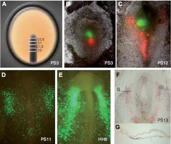
Fig. 1. Fate mapping of primitive streak cells. (A) Schematic diagram showing the four analyzed sites of the primitive streak (C0.5-C2). (B) Double microinjection of site C0.5 (CFSE, green spot) and C1.5 (CRSE, red spot), to show the rostro-lateral distribution of cells (C). Electroporated embryo at the level of segment C1.5 showing the subsequent distribution of cells at the level of the HFR (D) and then at the level of the two primitive endocardial tubes (E). Whole mount (F) and section (G) of an embryo subjected to double transplantation of sites C1 and C2 after ISH and double immunocytochemistry, showing in the mesoderm the expression of Usmaar (blue) and the distribution of cells from C2 (red), and in the endoderm the cells from C1 (brown).

прекардиальных клеток и участия организатора (рострального конца первичной полоски или Гензеновского узелка) в контроле и регуляции процесса кардиогенеза.

Results


A detailed picture was obtained of the location and movement of precardiac cells using a variety of techniques to construct a precise fate map. Also, with the design of different experimental procedures, we established several mechanisms involved in precardiac cells’ commitment and ability to differentiate when they are transplanted to ectopic locations, as well as shedding further light on the role of the organizer in the cardiogenic process.

Fate mapping of primitive streak cells


The location and movement of different groups of cells of the primitive streak during gastrulation was analyzed by means of double microinjections, double transplantation, and double immunocytochemistry. Simultaneously, we analyzed the expression of several genes related with precardiac areas using in situ hybridization (ISH). As seen in Fig. 1, four levels of the primitive streak (C0.5-C2) were subjected to double microinjection or transplantation, and subsequently identified by immunocytochemistry to show the pattern of migration through several locations in the developing chick, including precardiac and cardiac structures. Cells from the primitive streak were found to migrate bilaterally in


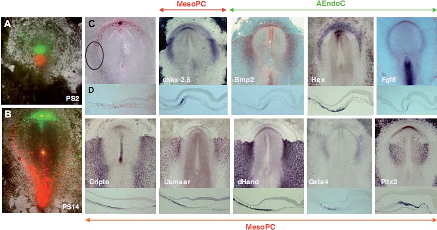
Fig. 2. Correlation between cells from sites C0.5 or C1.5, distributed in the HFR, and gene expression patterns. (A) Double microinjection of CFSE (green spot) and CRSE (red spot) to identify 12 hours later the rostro-lateral distribution of the cells (B). Double immunocytochemistry (C,D) shows the AEndoC (red) arriving from site C0.5, and the MesoPC (blue) from site C1.5. Note the overlap of the two groups of cells at the level of the HFR (ellipse). Whole mount and sections of embryos (PS14) subjected to ISH show at the level of the MesoPC the expression of cNkx-2.5, Cripto, Usmaar , dHand, GATA4, and Pitx2, and at the level of the AEndoC the expression of Bmp2, Hex, and Fgf8. A crescent-shaped pattern is observed in the expression of cNkx-2.5, Bmp2, and Hex, similar to the cell distribution observed after labeling the primitive streak cells.

a rostro-lateral direction, giving rise to the endodermal layer and mesodermal structures, and following a precise pattern of migration until their final location into the heart forming region (HFR) or primary cardiac field (PCF), where their differentiation takes place.
A few hours later, stages PS11-14 (Fig. 2), most cells located at the rostral site (C0.5) contributed in the anterior half of the embryo to forming the more anterior and lateral endodermal area, in a crescent-shaped pattern; while cells from a more caudal site (C1.5) at the same time contributed to forming bilateral mesodermal areas, reaching the border with the area pellucida, and overlapping with the caudal limits of the aforementioned crescent. At these stages, PS11-14, several specific markers are closely related to this cell distribution. A group of genes, including cNkx2.5 , Bmp2, and Hex, showed in whole mount a crescent of expression, similar to the cell distribution observed after the primitive streak cell labeling. At the level of the HFR (or PCF), we observed that cNkx-2.5 initially expressed at the level of the MesoPC, where there was expression of some other genes, including Cripto, Usmaar, dHand, GATA4, and Pitx2. The expression of Bmp2 and Hex, however, was at the level of the AEndoC, the location also observed for the expression of Fgf8.
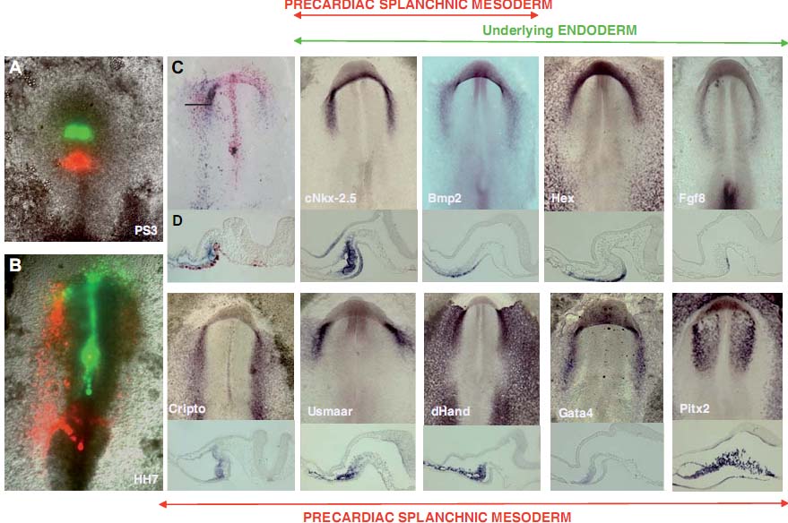
Analyzing the fate several hours later (HH7), we observed that most cells located at the rostral site (C0.5), which were to form the endodermal layer, contributed to forming the endoderm of the foregut, as well as the endoderm underlying the splanchnopleure (Fig. 3). Interestingly, we observed that the second group of cells of the primitive streak (C1.5) contributed cells to this bilateral precardiac splanchnic mesoderm, to form from HH7 the primitive endocardial tubes. Specific markers closely related to this step of cardiogenesis showed restricted expression at the level of the areas related with primitive endocardial tubes. Thus, from this stage, while cNkx-2.5 and Bmp2 were expressed in both the precardiac splanchnic mesoderm and the underlying endoderm, Cripto, Usmaar, dHand, GATA4, and Pitx2 expressed only in the former, and Hex and Fgf8 only in the latter.
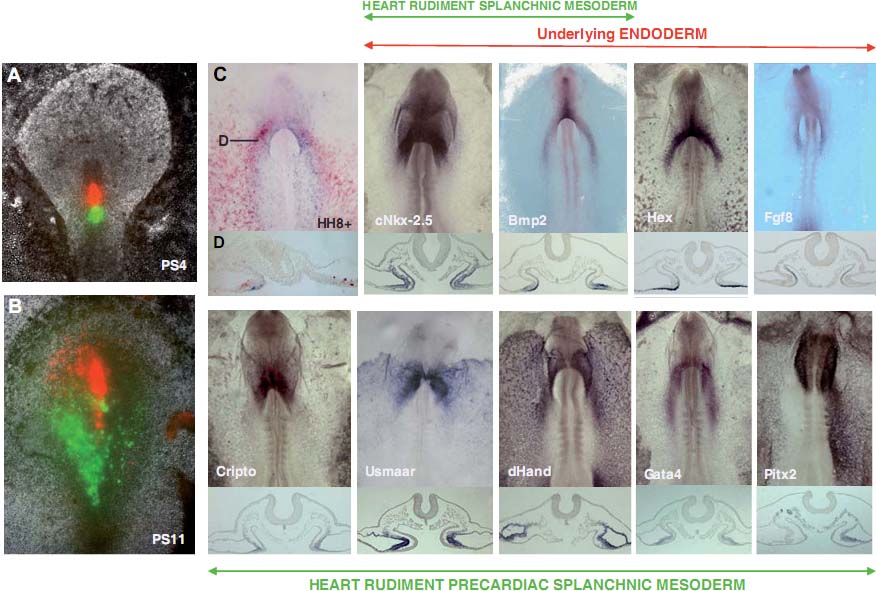
The study of another two groups of primitive streak cells, C1 and C2 (Fig. 4), located caudally to the foregoing sites, showed the first group of cells (coming from C1) to be distributed mainly at the level of the endoderm, underlying the second group of cells (coming from C2) located at the level of the splanchnic mesoderm of the heart rudiment, close to the amniocardiac vesicle portion of the coelom. From stage HH8, we followed the specific markers closely related to the process of cardiogenesis. At the level of the primitive endocardial tubes when they are beginning to fuse at the level of the midline, we observed expression of cNkx-2.5 and Bmp2 together with Cripto, Usmaar, dHand, GATA4, and GATA45-6 in the splanchnic mesoderm of heart rudiment, with the characteristic asymmetric expression of Pitx2 in the left lateral plate mesoderm. Moreover, Hex and Fgf8 expressed in the underlying endoderm together with cNkx-2.5 and Bmp2.
We observed that, when the two endocardial tubes finally move


Fig. 3. Correlation between cells from sites C0.5 or C1.5, distributed in the primitive endocardial tubes, and gene expression patterns. (A) Double microinjection of CFSE (green spot) and CRSE (red spot) to identify 14 hours later the rostro-lateral distribution of the cells (B). Double immunocytochemistry (C,D) shows the cells from site C1.5 forming the precardiac splanchnic mesoderm (blue), and cells from site C0.5 forming the underlying endoderm (red). Whole mount and sections of embryos (HH7-8) subjected to ISH show the expression of Cripto, Usmaar, dHand, GATA4, and Pitx2 at the level of the precardiac splanchnic mesoderm, and the expression of Hex and Fgf8 at the level of the underlying endoderm. Note that cNkx-2.5 and Bmp2 show expression in both layers.

to the midline and fuse to form the primitive heart tube, cells from the rostral-most site of the primitive streak mainly contribute to foregut endoderm and dorsal mesocardium, and the other more caudal sites analyzed form the myocardial and endocardial layers of the primitive heart tube (Fig. 5).
As in our previous fate maps, some cells from the head mesenchyme, blood vessels, ventral and dorsal aortae, and endoderm or mesoderm not involved in heart structures, were also observed in our experimental embryos.

Commitment and regulation mechanisms of precardiac cells


The state of commitment of precardiac cells located at the level of different segments of the primitive streak was studied by their transplantation into host embryos at different positions. We also studied the possible role of the rostral tip of the primitive streak as a potential regulatory mechanism in cardiogenesis.
Grafts of prospective cardiac cells into the germ cell crescent Groups of cells corresponding to sites C0.5 and C1 at stage PS3 when transplanted to the germ cell crescent at stage PS8 were able to form an endodermal-like layer, accompanied by a closely related tissue, which expressed the earliest specific cardiac marker cNkx-2.5 (Fig. 6A-D). Furthermore, groups of cells corresponding to sites C1.5 and C2 (Fig. 6E) were able to differentiate, expressing not only cNkx-2.5 but also specific markers of atrial, AMHC1 (Fig. 6F), and ventricular, VMHC1 (Fig. 6G,H), cardiac segments.
Grafts of prospective cardiac cells into prospective somitic sites, and vice versa Transplantation of cells (Fig. 7) from segment C1.5 of donor embryos at stage PS3 (precardiac cells) to site C1.5 at stage PS8 (presomitic site; Schoenwolf et al., 1992) showed the precardiac cells to change their fate, to form the somites. Furthermore, they expressed the specific somitic marker paraxis (Fig. 7B,C). Transplantation of cells from segment C1.5 of donor embryos at stage PS8 (presomitic cells) to site C1.5 at stage PS3 (precardiac site) showed the presomitic cells to also change their fate, to form


A Fig. 4 (Top). Correlation between cells from sites C1 or C2, distributed in the primitive endocardial tubes, and gene expression patterns. (A) Double microinjection of CRSE (red spot) and CFSE (green spot) to identify 16 hours later the rostro-lateral distribution of the cells (B). Double immunocytochemistry (C,D) shows the cells from site C2 forming the splanchnic mesoderm of the heart rudiment (red), and cells from site C1 forming the underlying endoderm (blue). Whole mount and sections of embryos (HH8-9) subjected to ISH show the expression of Cripto, Usmaar, dHand, GATA4, and Pitx2 at the level of the heart rudiment splanchnic mesoderm, and the expression of Hex and Fgf8 at the level of the underlying endoderm. Note that cNkx-2.5 and Bmp2 show expression in both layers.

heart, expressing cNkx-2.5 (Fig. 7D-G).
Grafts of the rostral tip of the primitive streak into HFR As can be seen in Fig. 8, the rostral tip of donor embryos at stage PS3 transplanted to HFR of a host embryo at stage PS11 gave rise a few hours later to an area of loss of expression of the early cardiac marker cNkx-2.5 at the level of the HFR in the host embryo (Fig. 8B). On the contrary, the Hensen’s node of donor embryos at stage PS8 transplanted to HFR (PS11) induced an increased area of expression of this marker (Fig. 8D).

Discussion


Одним из основных вопросов процесса кардиального развития является вопрос происхождения и поведения прекардиальных клеток до формирования органа. Известно (Lopez-Sanchez et al., 2001), что они обнаружимы уже на инициальных стадиях гаструляции (PS1) на уровне эпибласта и позднее во время их инвагинации через первичную полоску. Детальные карты судеб были сконструированы как для проспективных мезодермальных, так и энтодермальных клеток (Schoenwolf et al., 1992; Sawada and Aoyama, 1999; Lawson and Schoenwolf, 2003; Kimura et al., 2006; Chapman et al ., 2007), однако они не были сфокусированы на происхождении и замещении проспективных кардиальных клеток во время развития сердца. Хотя разные исследователи (Fishman and Chien, 1997; Ehrman and Yutzey, 1999; Redkar et al., 2001) внесли вклад в конструирование карты судеб прекардиальных клеток, всё ещё оставались некоторые аспекты, нуждающиеся в объяснении, такие как корреляция между положение прекардиальных клеток на уровне первичной полоски с их расположением на уровне HFR (или PCF), и их финальной позицией в примитивной сердечной трубке. В настоящем исследовании мы провели детальный анализ происхождения, расположения и перемещения прекардиальных клеток вплоть до момента, когда их судьба детерминировалась к формированию структуры HFR, эндокардиальных трубок и наконец, примитивной сердечной трубки. Наши результаты показали. что наиболее ростральный регион первичной полоски, за исключением Гензеновского узелка, мигрирует билатерально в ростро-латеральном направлении, чтобы сформировать часть преимущественно энтодермального слоя, включая AEndoC. Клетки первичной полоски, расположенные непосредственно каудальнее этого региона. следуют параллельно, но в более латеральном направлении и составляют MesoPC, которые позднее становятся прекардиальной спланхнической мезодермой. Это может быть очень важным аспектом, принимая во внимание кардиогенез, т.к. было предположено (Schultheiss et al., 1995, 1997; Ehrman and Yutzey, 1999) , что the AEndoC индуцируют инициацию кардиогенеза, действуя на MesoPC. Было предположено, что клетки, которые инвагинируют на уровне наиболее ростральных сегментов первичной полоски играют роль и на более поздних стадиях в индукции и регуляции дифференцировки клеток, которые инвагинируют через каудальные сегменты и которые формируют слои, характерные для структур примитивной сердечной трубки (Fig. 9). Ранее нами было установлено (Schoenwolf et al., 1992; Garcia-Martinez and Schoenwolf, 1993), что наиболее ростральные клетки первичной полоски в основном формируют артериальный полюс сердечной трубки, а более каудальные в основном венозный полюс и что даже на ст. PS8 некоторые клетки Гензеновского узелка способны формировать часть эндокарда артериального полюса (Schoenwolf et al., 1992; Kirby et al., 2003). По результатом большой группы техник, использованных в данном исследовании, подтверждена гипотеза, что клетки первичной полоски состоят из двух групп разного значения. Одна группа клеток, расположенная преимущественно в более ростральной области первичной полоски (C05-C1), может быть охарактеризована по своей проспективной способности индукции и регуляции (AEndoC) второй группы клеток. Эта вторая группа, расположенная преимущественно в более каудальных сегментах (C1.5-C2.5), предназначена сформировать и дифференцировать MesoPC, чтобы создать прекардиальную спланхническую мезодерму и затем слои примитивной сердечной трубки (Fig. 9).
Разные исследователи (Fishman and Chien, 1997; Ehrman and Yutzey, 1999; Redkar et al., 2001) пытались установить конфигурацию и расположение клеток, составляющих HFR, и ограничить эту область, используя технику DiI crystal имплантаций и DiI инъекций. В результате появились доказательства расхождений между этими исследованиями в отношении границ и организации прекардиальных клеток в HFR, т.к. отсутствуют четкие анатомические границы, определяющие этот регион. В попытке внести вклад в понимание процесса образования, разграничения и миграции клеток в HFR, мы здесь описали исследование картирования судеб прекардиальных клеток первичной полоски вместе с детальным анализом экспрессии разных специфических генов сердца или интимно связанных с его развитием. Наши результаты показали, что прекардиальные клетки от их местоположения в первичной полоске до их расположения в примитивной сердечной трубке, и клетки интимно связанные с ними (AEndoC), характеризуются присутствием иерархическоео паттерна экспрессии разных генов. Экспрессия Bmp2, который считается индуктором кардиогенеза (Lough et al., 1996; Schultheiss et al., 1997; Andree et al., 1998; Schlange et al., 2000; Alsan and Schultheiss, 2002; Lopez-Sanchez et al., 2002) начинается в AEndoC, которые считаются первой группой клеток, прибывших из ростральных сайтов первичной полоски, а спустя несколько часов обнаруживается как в прекардиальной спланхнической мезодерме, так и в подлежащей энтодерме. Однако самый ранний кардиальный маркёр cNkx-2.5 (Schultheiss et al., 1995) впервые экспрессируется на уровне MesoPC, чти клетки происходят из более каудальных сегментов первичной полоски, что сопровождается позднее экспрессией на уровне MesoPC и AEndoC одновременно. С точки зрения этих данных и анализа генной экспрессии на уровне первичной полоски на этих стадиях (PS1- 14, Lopez-Sanchez et al., 2005), нами была высказана гипотеза, что вторая группа клеток первичной полоски (проспективные MesoPC) регулируется первой группой (проспективные AEndoC), которая, в свою очередь, регулируется при посредстве рострального кончика Гензеновского узелка (Fig. 9). Более того, этот процесс инициации регуляции кардиогенеза д. контролироваться генами, экспрессия которых необходима на уровне AEndoC, такими как Fgf8 (Alsan and Schultheiss, 2002) и Hex (Zhang et al., 2004), тогда как др. гены. такие как Cripto (Colas and Schoenwolf, 2000), Usmaar (Colas et al., 2000), dHand (Srivastava et al., 1995), GATA4, GATA4-5-6
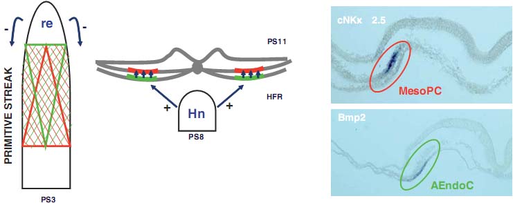


Fig. 9. A model of the significance of primitive streak precardiac cells. The precardiac cells of the primitive streak are distributed into two groups with different significance. The group of cells located in the more rostral sites of the primitive streak, with a decreasing gradient towards more caudal sites (green triangle), is regulated by the rostral end (re), and is responsible for the formation of the endoderm layer (green line) characterized by the expression of specific genes, such as the initial expression of Bmp2 (green ellipse). The second group is located in more caudal sites of the primitive streak, with a decreasing gradient towards the more rostral sites (red triangle). This group will be responsible for the formation of the MesoPC (red line), differentiating subsequently to form the precardiac splanchnic mesoderm and primitive heart tube. It is regulated by the cells of the previous group, and expresses specific genes, such as the initial expression of cNkx-2.5 (red ellipse). At later stages, the Hensen's node (Hn) regulates the cell differentiation of the HFR.

(Laverriere et al., 1994; Jiang et al., 1998) и Pitx2 (Piedra et al., 1998), д. регулироваться, чтобы экспрессироваться в MesoPC и поддерживать свою экспрессию в ходе дифференцировки этих клеток в направлении спланхнической мезодермы и примитивных эндокардиальных трубок. Некоторые др. необходимы на более поздних фазах кардиального морфогенеза, включая AMHC1 (Yutzey et al. , 1994; Somi et al., 2006), VMHC1 (Bisaha and Bader, 1991; Somi et al., 2006), Tbx1, (Huynh et al., 2007) и Tbx5 (Bruneau et al., 1999; Plageman and Yutzey, 2004).
Итак эти данные показывают, что потенциал клеток первичной полоски дифференцироваться в направлении сердца д. зависеть не только от их собственных пространственно-временных характеристик, но и также от индуктивных и/или регуляторных факторов, с которыми они поддерживают интимные взаимоотношения. По этой причине, в данной работе мы анализировали следующие два аспекта: детерминация прекардиальных клеток первичной полоски и их регуляторные механизмы. Наши результаты показали, что наиболее ростральные прекардиальные клетки первичной полоски, трансплантированные в серп зародышевых клеток, способны давать энтодерма-подобный слой, сопровождаемый близко родственной тканью, которая экспрессирует самый ранний кардиальный маркер cNkx-2.5, тогда как более каудальные клетки обладали способностью дифференцировки и экспрессии cNkx-2.5, а также маркеров кардиальных секторов, AMHC1 и VMHC1. Эти данные указывают на то, что прекардиальные клетки первичной полоски уже обладают информацией о своей судьбе в отношении кардиогенеза и подтверждают гипотезу, предложенную выше (Fig. 9). Тем не менее, если прекардиальные клетки первичной полоски гомотипически или гетеротипически трансплантировать в пресомитные сайты первичной полоски, то они теряют свою способность мигрировать в направлении сердца и экспрессии ими специфических кардиальных генов (и vice versa). Эти данные подтверждают гипотезу, что на уровне первичной полоски существуют факторы, которые детерминируют механизмы становления регуляции перемещения и дифференцировки её клеток, со способностью модифицировать их инициальную информацию. В своей попытке получить новые данные относительно возможных факторов, участвующих в этом процессе индукции и регуляции, мы оказались способны продемонстрировать участие рострального кончика первичной полоски, показав. то этот конец на стадии PS2-5 первичной полоски негативно регулирует экспрессию генов, участвующих в инициации кардиогенеза. Свойства Гензеновского узелка на поздних стадиях с разными морфологическими и молекулярными характеристиками (Lopez-Sanchez et al., 2005), необходимы для начала этого процесса. Эти находки подтверждают наши более ранние результаты (Lopez-Sanchez et al., 2002) о способности Гензеновского узелка обеспечивать индукцию процесса кардиогенеза.
Исследования по картированию судеб и анализ способности к дифференцировке и/или индукции клеток первичной полоски недостаточны, чтобы иметь полное представление о факторах, которые детерминируют процесс кардиогенеза и кардиального морфогенеза. Необходим также экспериментальный анализ молекулярных факторов, которые регулируют MesoPC и AEndoC, которые возникают на уровне первичной полоски.
Сайт создан в системе uCoz